Weight-Bearing CT Scanner

CurveBeam HiRise

HiRise — bilateral upright CT, 20 cm × 40 cm field of view, 0.3 mm slices, approx. 26 s scan, 205 kg capacity

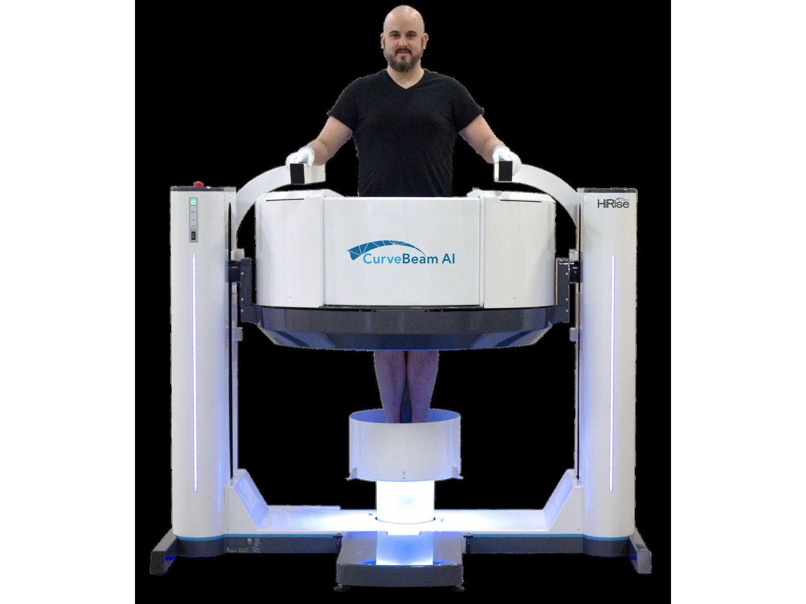
CurveBeam HiRise from CurveBeam is a bilateral weight-bearing CT scanner for upright 3D imaging of the lower extremities under natural load, with 0.3 mm slice thickness and a scan time of approx. 26 seconds.

Manufactured byCurveBeam
CurveBeam logo
CurveBeam HiRise weight-bearing ct scanner
CurveBeam HiRise from CurveBeam.
20 × 40 cmField of view (H × Ø)
0.3 mmSlice thickness
approx. 26 sScan time
205 kgPatient weight capacity
CE · FDAApprovals
Overview

Upright 3D CT of the lower extremities under load

HiRise is a weight-bearing CT scanner developed by CurveBeam AI for upright 3D imaging of the lower extremities under natural load. Patients stand during scanning, which shows anatomical alignment under load in a way that supine CT cannot replicate.

The large field of view captures both limbs simultaneously, enabling bilateral assessment of foot, ankle, knee and hip structures. The system supports a patient weight of up to 450 lbs (205 kg). With 0.3 mm slice thickness and a scan time of approx. 26 seconds, HiRise produces high-resolution volumetric datasets for surgical planning, deformity assessment and post-operative evaluation.

HiRise is intended for orthopedic clinics, imaging centers and specialty practices that need a dedicated upright CT system for weight-bearing biomechanical evaluation.

Applications

Where the HiRise is used

HiRise is used where weight-bearing 3D CT imaging of the foot, ankle, knee and lower extremity is required.

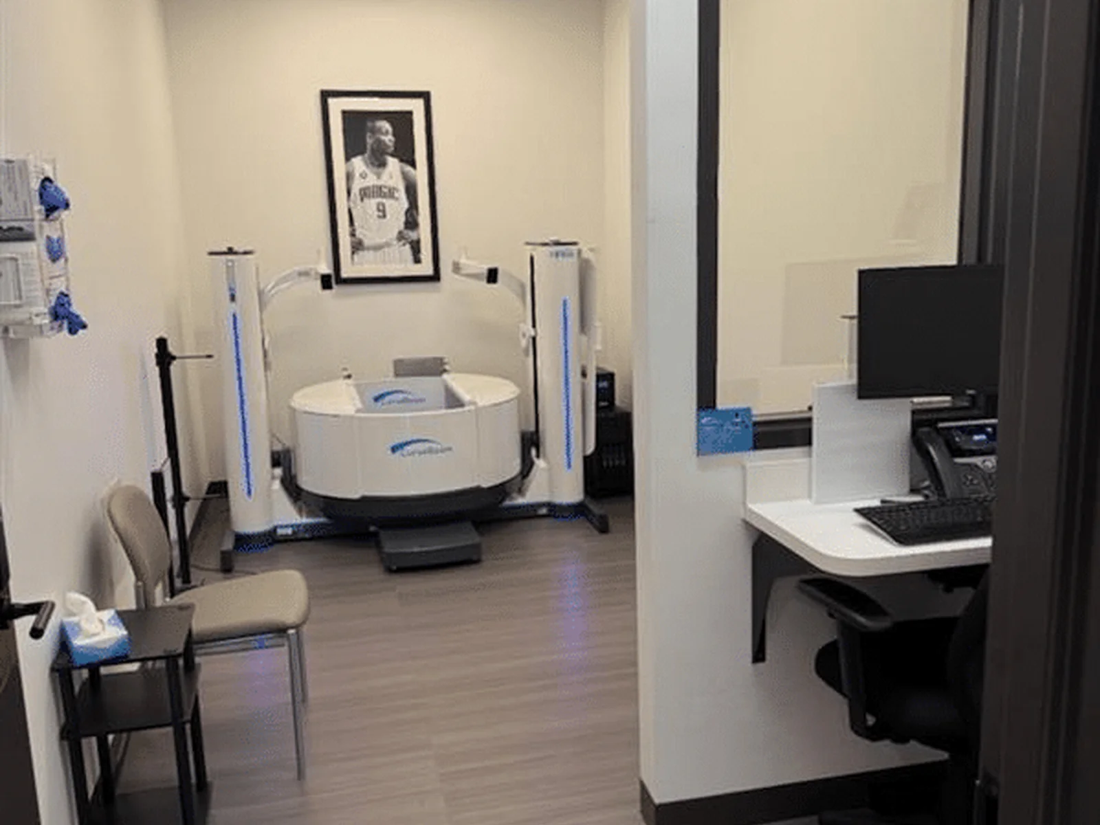
CurveBeam HiRise in use – orthopedic clinics
Orthopedics

Orthopedic Clinics

Weight-bearing CT for deformity assessment and surgical planning.

Podiatry

Podiatry Centers

Upright 3D imaging of the foot and ankle under load.

Sports Medicine

Sports-Medicine Facilities

Lower-extremity evaluation under natural load.

Imaging

Imaging Departments

Dedicated weight-bearing 3D CT for lower-extremity evaluation.

Orthopedic clinicsPodiatry centersSports medicineImaging departmentsFoot and ankleKnee evaluation
Features

Weight-bearing CT, coverage and speed

Weight-Bearing Imaging

  • Upright, weight-bearing 3D CT imaging.
  • Bilateral imaging of the lower extremities under natural load.
  • Designed for orthopedic and biomechanical assessment.

Field of View & Capacity

  • Large field of view: 20 cm height × 40 cm diameter.
  • Patient weight capacity: 450 lbs (205 kg).

Resolution & Speed

  • Sub-millimeter slice thickness of 0.3 mm.
  • Scan time of approx. 26 seconds.
  • High-resolution volumetric reconstruction.
Benefits

Why it suits orthopedic assessment

Alignment Under Load. Shows anatomical alignment under weight-bearing conditions.
Wide Patient Range. The high weight capacity accommodates a wide range of patients.
Spatial Resolution. High spatial resolution improves diagnostic confidence.
Fast Scanning. The short scan time supports patient comfort and workflow efficiency.
Bilateral Comparison. Bilateral imaging supports comparative assessment of deformity and alignment.
Surgical Planning. Suited to orthopedic diagnostics and surgical planning.

Need support with this weight-bearing CT scanner?

Share your clinical department, main lower-extremity applications, available room space and expected patient volume, and our medical technologies team will support you with the suitable product information.

Request Information
Medical Technologies Division

Request Medical Technologies Information

Share your requirements — our specialists reply within one working day.

Noor Scientific & Trade

Request Information

Share your requirements — our team will route your inquiry to the right division and reply within one working day.